


Image

The Neurosciences department at Apollo Hospitals, Gurgaon treats disorders of the brain, spinal cord, nerves and muscles — stroke, epilepsy, headache, Parkinson's disease, brain and spine tumours, nerve and muscle disease, head and spinal injury, and conditions affecting memory and movement. Neurology and neurosurgery work as a single unit, alongside neurocritical care, neurointervention and rehabilitation.
Emergency assessment, CT and MRI imaging, the catheterisation laboratory, operating theatres, a neuro intensive care unit and therapy services are all within the same hospital. That concentration is what distinguishes leading neurosciences care in Gurgaon, because outcomes in conditions such as stroke, head injury and spinal cord compression depend directly on how fast a patient moves from arrival to imaging to treatment.
Most cases are reviewed jointly. A brain tumour may involve a neurosurgeon, a neuroradiologist, an oncologist and a pathologist before the approach is fixed; a patient with epilepsy that has not responded to medication may be assessed by a neurologist and a neurosurgeon together. Care continues after the acute phase through rehabilitation and long-term follow-up. Treatment plans differ from patient to patient and are decided after examination and investigation.
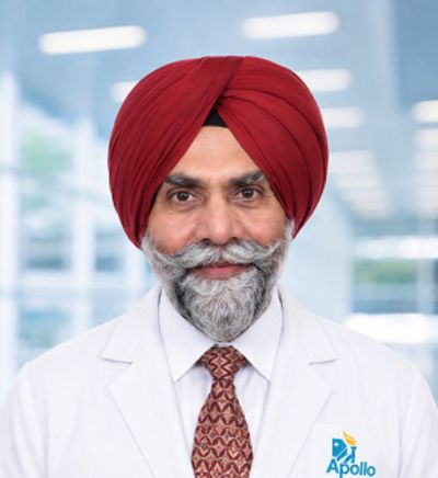
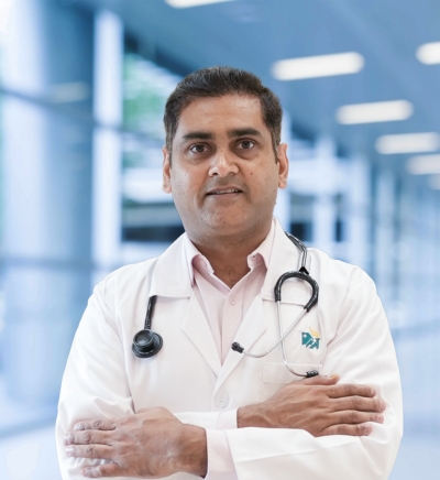
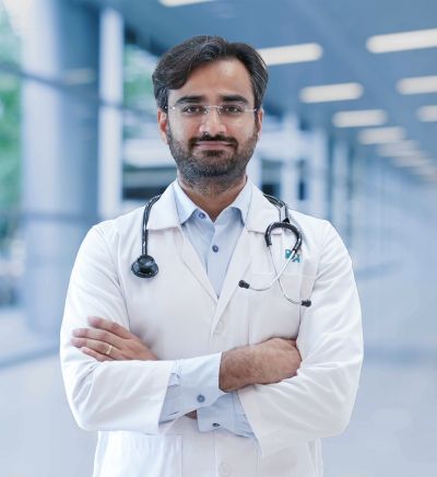
Brain, spine and nerve care at Apollo Hospitals, Gurgaon is provided by neurologists, neurosurgeons, spine surgeons, neurointerventionists and neurocritical care specialists, supported by neuroradiologists, neuroanaesthetists, physiotherapists, occupational and speech therapists and clinical psychologists. Complex cases — tumours, aneurysms, drug-resistant epilepsy, movement disorders considered for surgery — are discussed jointly across the medical and surgical teams rather than managed by one specialist alone. Our specialists include:

A detailed, radiation-free scan using a magnetic field, giving the clearest available view of brain tissue, the spinal cord and nerves. You lie still inside the scanner for 20 to 45 minutes and the machine is noisy, so earplugs are provided. Tell the team beforehand about any pacemaker, implant or metal in the body.
Imaging of the brain's blood vessels using contrast, which maps narrowing, aneurysms and malformations without a catheter.
Small sensors placed on the scalp record the brain's electrical activity. It is completely painless, involves no needles and takes 30 to 60 minutes. It is used mainly in the assessment of seizures.
A needle inserted into the lower back under local anaesthesia collects a small sample of the fluid surrounding the spinal cord. It takes 15 to 30 minutes and is essential in suspected meningitis, encephalitis and certain inflammatory conditions. Headache afterwards is the common side effect and usually settles with rest and fluids.
Painless ultrasound assessment of blood flow in the neck and brain arteries, used after stroke and TIA to identify narrowing that may need treatment.
Structured tests of memory, attention, language and problem-solving administered by a psychologist over one or more sessions. They clarify the pattern and severity of cognitive change and are not pass-or-fail.
Most neurological conditions are treated medically. Surgery and catheter-based procedures are used where they offer clear benefit, and the choice is made jointly between the neurology and neurosurgery teams.
Clot-dissolving medication given intravenously within the accepted time window, and mechanical thrombectomy — removal of the clot through a catheter threaded up to the brain artery — in selected patients with a large vessel blockage. Both are followed by care in a stroke or neuro intensive care unit.
Antiplatelet or anticoagulant medication, blood pressure and cholesterol control, diabetes management, and procedures to treat carotid narrowing where indicated.
Anti-seizure medication selected according to seizure type, age, sex and other conditions, with doses adjusted until seizures are controlled with acceptable side effects. Most patients are managed on a single medicine.
Acute medication for attacks, preventive treatment where attacks are frequent, identification and withdrawal of overused painkillers, and botulinum toxin for chronic migraine in appropriate patients.
Implantation of fine electrodes into specific brain areas, connected to a pulse generator placed under the skin, used in Parkinson's disease, tremor and dystonia that are no longer adequately controlled by medication.


Sudden facial drooping on one side, weakness or numbness in an arm or leg, slurred or confused speech, sudden loss of balance or vision. Note the time symptoms started and go to a hospital with stroke facilities immediately or call an ambulance. Do not wait to see whether it improves, do not give food or water, and do not drive yourself. Treatment options narrow with every hour that passes.
It depends on the type of stroke and the imaging findings. Clot-dissolving medication is generally given within about four and a half hours of symptom onset. Clot removal through a catheter can help selected patients considerably later, guided by specialised scans. Arriving early gives access to the widest range of options. [VERIFY time windows against current national guidance]
A neurologist diagnoses and treats nervous system conditions with medication and other non-surgical approaches. A neurosurgeon operates on the brain, spine and nerves. Many patients see a neurologist first, and surgery is involved only if it is needed. Both work together on cases where the choice is not clear-cut.
It is painless and uses no radiation. The scanner is noisy and enclosed, which some people find uncomfortable, and mild sedation can be arranged for those who are claustrophobic. You must inform the team about any pacemaker, implanted device, metal fragments or cochlear implant, as some are unsafe in the magnetic field.
No. An EEG only records the brain's own electrical activity through sensors stuck to the scalp. Nothing is passed into the body, and it is entirely painless. You may be asked to breathe deeply or look at a flashing light during the test.
Most people with epilepsy achieve good seizure control on medication. Whether treatment is lifelong depends on the type of epilepsy, the cause and how long seizures have been absent — in some cases medication can be withdrawn gradually after a sustained seizure-free period, under specialist supervision. Never stop anti-seizure medication on your own.



 Best Hospital Near me Chennai
Best Hospital Near me Bangalore
Best Hospital Near me Hyderabad
Best Hospital Near me Mumbai
Best Hospital Near me Delhi
Best Hospital Near me Kolkata
Best Hospital Near me Pune
Best Hospital Near me Ahmedabad
Best Hospital Near me Bhubaneswar
Best Hospital Near me Bilaspur
Best Hospital Near me Guwahati
Best Hospital Near me Indore
Best Hospital Near me Kochi
Best Hospital Near me Lucknow
Best Hospital Near me Madurai
Best Hospital Near me Mysore
Best Hospital Near me Nashik
Best Hospital Near me Noida
Best Hospital Near me Rourkela
Best Hospital Near me Visakhapatnam
Cardiologist Near me in Chennai
Cardiologist Near me in Bangalore
Cardiologist Near me in Hyderabad
Cardiologist Near me in Mumbai
Cardiologist Near me in Delhi
Cardiologist Near me in Kolkata
Cardiologist Near me in Pune
Cardiologist Near me in Ahmedabad
Cardiologist Near me in Bhubaneswar
Cardiologist Near me in Bilaspur
Cardiologist Near me in Guwahati
Cardiologist Near me in Indore
Cardiologist Near me in Kochi
Cardiologist Near me in Lucknow
Cardiologist Near me in Madurai
Cardiologist Near me in Mysore
Cardiologist Near me in Nashik
Cardiologist Near me in Noida
Cardiologist Near me in Rourkela
Cardiologist Near me in Visakhapatnam
Neurologist Near me in Chennai
Neurologist Near me in Bangalore
Neurologist Near me in Hyderabad
Neurologist Near me in Mumbai
Neurologist Near me in Delhi
Neurologist Near me in Kolkata
Neurologist Near me in Ahmedabad
Neurologist Near me in Bhubaneswar
Neurologist Near me in Bilaspur
Neurologist Near me in Guwahati
Neurologist Near me in Indore
Neurologist Near me in Kochi
Neurologist Near me in Lucknow
Neurologist Near me in Madurai
Neurologist Near me in Mysore
Neurologist Near me in Nashik
Neurologist Near me in Noida
Neurologist Near me in Rourkela
Neurologist Near me in Visakhapatnam
Orthopedician Near me in Chennai
Orthopedician Near me in Bangalore
Orthopedician Near me in Hyderabad
Orthopedician Near me in Mumbai
Orthopedician Near me in Delhi
Orthopedician Near me in Kolkata
Orthopedician Near me in Pune
Orthopedician Near me in Ahmedabad
Orthopedician Near me in Bhubaneswar
Orthopedician Near me in Bilaspur
Orthopedician Near me in Guwahati
Orthopedician Near me in Indore
Orthopedician Near me in Kochi
Orthopedician Near me in Lucknow
Orthopedician Near me in Madurai
Orthopedician Near me in Mysore
Orthopedician Near me in Nashik
Orthopedician Near me in Noida
Orthopedician Near me in Rourkela
Orthopedician Near me in Visakhapatnam
Oncologist Near me in Chennai
Oncologist Near me in Bangalore
Oncologist Near me in Hyderabad
Oncologist Near me in Mumbai
Oncologist Near me in Delhi
Oncologist Near me in Kolkata
Oncologist Near me in Pune
Oncologist Near me in Ahmedabad
Oncologist Near me in Bhubaneswar
Oncologist Near me in Bilaspur
Oncologist Near me in Guwahati
Oncologist Near me in Indore
Oncologist Near me in Kochi
Oncologist Near me in Lucknow
Oncologist Near me in Madurai
Oncologist Near me in Mysore
Oncologist Near me in Nashik
Oncologist Near me in Noida
Oncologist Near me in Rourkela
Gastroenterologist Near me in Chennai
Gastroenterologist Near me in Bangalore
Gastroenterologist Near me in Hyderabad
Gastroenterologist Near me in Mumbai
Gastroenterologist Near me in Delhi
Gastroenterologist Near me in Kolkata
Gastroenterologist Near me in Pune
Gastroenterologist Near me in Ahmedabad
Gastroenterologist Near me in Bhubaneswar
Gastroenterologist Near me in Bilaspur
Gastroenterologist Near me in Guwahati
Gastroenterologist Near me in Indore
Gastroenterologist Near me in Kochi
Gastroenterologist Near me in Lucknow
Gastroenterologist Near me in Madurai
Gastroenterologist Near me in Mysore
Gastroenterologist Near me in Nashik
Gastroenterologist Near me in Noida
Gastroenterologist Near me in Rourkela
Gastroenterologist Near me in Visakhapatnam
Transplant Surgeon Near me in Chennai
Transplant Surgeon Near me in Bangalore
Transplant Surgeon Near me in Hyderabad
Transplant Surgeon Near me in Mumbai
Transplant Surgeon Near me in Delhi
Transplant Surgeon Near me in Kolkata
Transplant Surgeon Near me in Indore
Transplant Surgeon Near me in Kochi
Transplant Surgeon Near me in Lucknow
Transplant Surgeon Near me in Mysore
ENT Specialist Near me in Chennai
ENT Specialist Near me in Bangalore
ENT Specialist Near me in Hyderabad
ENT Specialist Near me in Mumbai
ENT Specialist Near me in Delhi
ENT Specialist Near me in Kolkata
ENT Specialist Near me in Pune
ENT Specialist Near me in Ahmedabad
ENT Specialist Near me in Bhubaneswar
ENT Specialist Near me in Bilaspur
ENT Specialist Near me in Guwahati
ENT Specialist Near me in Indore
ENT Specialist Near me in Kochi
ENT Specialist Near me in Lucknow
ENT Specialist Near me in Madurai
ENT Specialist Near me in Mysore
ENT Specialist Near me in Nashik
ENT Specialist Near me in Noida
ENT Specialist Near me in Rourkela
ENT Specialist Near me in Visakhapatnam
Pulmonologist Near me in Chennai
Pulmonologist Near me in Bangalore
Pulmonologist Near me in Hyderabad
Pulmonologist Near me in Mumbai
Pulmonologist Near me in Delhi
Pulmonologist Near me in Kolkata
Pulmonologist Near me in Ahmedabad
Pulmonologist Near me in Bhubaneswar
Pulmonologist Near me in Bilaspur
Pulmonologist Near me in Guwahati
Pulmonologist Near me in Indore
Pulmonologist Near me in Kochi
Pulmonologist Near me in Lucknow
Pulmonologist Near me in Madurai
Pulmonologist Near me in Mysore
Pulmonologist Near me in Nashik
Pulmonologist Near me in Noida
Pulmonologist Near me in Rourkela
Pulmonologist Near me in Visakhapatnam
Dentist Near me in Chennai
Dentist Near me in Bangalore
Dentist Near me in Hyderabad
Dentist Near me in Delhi
Dentist Near me in Kolkata
Dentist Near me in Ahmedabad
Dentist Near me in Bhubaneswar
Dentist Near me in Bilaspur
Dentist Near me in Guwahati
Dentist Near me in Kochi
Dentist Near me in Lucknow
Dentist Near me in Madurai
Dentist Near me in Nashik
Dentist Near me in Noida
Dentist Near me in Rourkela
Dentist Near me in Visakhapatnam
Pediatrician Near me in Chennai
Pediatrician Near me in Bangalore
Pediatrician Near me in Hyderabad
Pediatrician Near me in Mumbai
Pediatrician Near me in Delhi
Pediatrician Near me in Kolkata
Pediatrician Near me in Pune
Pediatrician Near me in Ahmedabad
Pediatrician Near me in Bhubaneswar
Pediatrician Near me in Bilaspur
Pediatrician Near me in Guwahati
Pediatrician Near me in Indore
Pediatrician Near me in Kochi
Pediatrician Near me in Lucknow
Pediatrician Near me in Madurai
Pediatrician Near me in Mysore
Pediatrician Near me in Nashik
Pediatrician Near me in Noida
Pediatrician Near me in Visakhapatnam
Dermatologist Near me in Chennai
Dermatologist Near me in Bangalore
Dermatologist Near me in Hyderabad
Dermatologist Near me in Delhi
Dermatologist Near me in Kolkata
Dermatologist Near me in Ahmedabad
Dermatologist Near me in Bhubaneswar
Dermatologist Near me in Bilaspur
Dermatologist Near me in Guwahati
Dermatologist Near me in Indore
Dermatologist Near me in Kochi
Dermatologist Near me in Lucknow
Dermatologist Near me in Madurai
Dermatologist Near me in Mysore
Dermatologist Near me in Noida
Dermatologist Near me in Visakhapatnam
Urologist Near me in Chennai
Urologist Near me in Bangalore
Urologist Near me in Hyderabad
Urologist Near me in Delhi
Urologist Near me in Kolkata
Urologist Near me in Pune
Urologist Near me in Ahmedabad
Urologist Near me in Bhubaneswar
Urologist Near me in Bilaspur
Urologist Near me in Guwahati
Urologist Near me in Indore
Urologist Near me in Kochi
Urologist Near me in Lucknow
Urologist Near me in Madurai
Urologist Near me in Mysore
Urologist Near me in Nashik
Urologist Near me in Noida
Urologist Near me in Rourkela
Urologist Near me in Visakhapatnam
Endocrinologist and Diabetologist Near me in Chennai
Endocrinologist and Diabetologist Near me in Bangalore
Endocrinologist and Diabetologist Near me in Hyderabad
Endocrinologist and Diabetologist Near me in Mumbai
Endocrinologist and Diabetologist Near me in Delhi
Endocrinologist and Diabetologist Near me in Kolkata
Endocrinologist and Diabetologist Near me in Pune
Endocrinologist and Diabetologist Near me in Ahmedabad
Endocrinologist and Diabetologist Near me in Bhubaneswar
Endocrinologist and Diabetologist Near me in Bilaspur
Endocrinologist and Diabetologist Near me in Guwahati
Endocrinologist and Diabetologist Near me in Indore
Endocrinologist and Diabetologist Near me in Kochi
Endocrinologist and Diabetologist Near me in Lucknow
Endocrinologist and Diabetologist Near me in Madurai
Endocrinologist and Diabetologist Near me in Mysore
Endocrinologist and Diabetologist Near me in Noida
Endocrinologist and Diabetologist Near me in Rourkela
Endocrinologist and Diabetologist Near me in Visakhapatnam
Gynecologist Near me in Chennai
Gynecologist Near me in Bangalore
Gynecologist Near me in Hyderabad
Gynecologist Near me in Mumbai
Gynecologist Near me in Delhi
Gynecologist Near me in Kolkata
Gynecologist Near me in Ahmedabad
Gynecologist Near me in Bhubaneswar
Gynecologist Near me in Bilaspur
Gynecologist Near me in Guwahati
Gynecologist Near me in Indore
Gynecologist Near me in Kochi
Gynecologist Near me in Lucknow
Gynecologist Near me in Madurai
Gynecologist Near me in Mysore
Gynecologist Near me in Nashik
Gynecologist Near me in Noida
Gynecologist Near me in Rourkela
Gynecologist Near me in Visakhapatnam
General Physician Near me in Chennai
General Physician Near me in Bangalore
General Physician Near me in Hyderabad
General Physician Near me in Mumbai
General Physician Near me in Delhi
General Physician Near me in Kolkata
General Physician Near me in Pune
General Physician Near me in Ahmedabad
General Physician Near me in Bhubaneswar
General Physician Near me in Bilaspur
General Physician Near me in Guwahati
General Physician Near me in Indore
General Physician Near me in Kochi
General Physician Near me in Lucknow
General Physician Near me in Madurai
General Physician Near me in Mysore
General Physician Near me in Nashik
General Physician Near me in Noida
General Physician Near me in Visakhapatnam
Psychologist Near me in Chennai
Psychologist Near me in Bangalore
Psychologist Near me in Hyderabad
Psychologist Near me in Mumbai
Psychologist Near me in Delhi
Psychologist Near me in Kolkata
Psychologist Near me in Ahmedabad
Psychologist Near me in Bhubaneswar
Psychologist Near me in Bilaspur
Psychologist Near me in Guwahati
Psychologist Near me in Indore
Psychologist Near me in Kochi
Psychologist Near me in Lucknow
Psychologist Near me in Madurai
Psychologist Near me in Mysore
Psychologist Near me in Nashik
Psychologist Near me in Noida
Psychologist Near me in Visakhapatnam
General Surgeon Near me in Chennai
General Surgeon Near me in Bangalore
General Surgeon Near me in Hyderabad
General Surgeon Near me in Mumbai
General Surgeon Near me in Delhi
General Surgeon Near me in Kolkata
General Surgeon Near me in Pune
General Surgeon Near me in Ahmedabad
General Surgeon Near me in Bhubaneswar
General Surgeon Near me in Bilaspur
General Surgeon Near me in Guwahati
General Surgeon Near me in Indore
General Surgeon Near me in Lucknow
General Surgeon Near me in Madurai
General Surgeon Near me in Mysore
General Surgeon Near me in Nashik
General Surgeon Near me in Noida
General Surgeon Near me in Rourkela
Best Hospital in Karukutty, Cochin
Best Hospital in Greams Road, Chennai
Best Hospital in Kuvempunagar, Mysore
Best Hospital in Vanagaram, Chennai
Best Hospital in Teynampet, Chennai
Best Hospital in OMR, Chennai
Best Cancer Hospital in Bhat, Gandhinagar, Ahmedabad
Best Cancer Hospital in Electronic City, Bangalore
Best Cancer Hospital in Teynampet, Chennai
Best Cancer Hospital in HSR Layout, Bangalore
Best Proton Cancer Centre in Chennai
Best Children's Hospital in Thousand Lights, Chennai
Best Women’s Hospital in Thousand Lights, Chennai
Best Hospital in Paschim Boragaon, Guwahati
Best Hospital in P H Road, Chennai
Best Heart Centre in Thousand Lights, Chennai
Best Hospital in Jubilee Hills, Hyderabad
Best Hospital in Tondiarpet, Chennai
Best Hospital in Kotturpuram, Chennai
Best Hospital in Kovai Road, Karur
Best Hospital in Karapakkam, Chennai
Best Hospital in Arilova, Vizag
Best Hospital in Kanpur Road, Lucknow
Best Hospital in Sector-26, Noida
Best Hospital in Gandhinagar, Ahmedabad
Best Hospital in Aragonda, Andhra Pradesh
Best Hospital in Bannerghatta Road, Bangalore
Best Hospital in Unit-15, Bhubaneswar
Best Hospital in Seepat Road, Bilaspur
Best Hospital in Ellisbridge, Ahmedabad
Best Hospital in New Delhi
Best Hospital in DRDO, Hyderabad
Best Hospital in G S Road, Guwahati
Best Hospital in Hyderguda, Hyderabad
Best Hospital in Vijay Nagar, Indore
Best Hospital in Suryaraopeta Main Road, Kakinada
Best Hospital in Canal Circular Road, Kolkata
Best Hospital in CBD Belapur, Navi Mumbai
Best Hospital in Panchavati, Nashik
Best Hospital in secunderabad, Hyderabad
Best Hospital in Seshadripuram, Bangalore
Best Hospital in Waltair Main Road, Visakhapatnam
Best Hospital in Subhash Nagar Road, Karimnagar
Best Hospital in Managari, Karaikudi
Best Hospital in Arepally, Warangal
Best Hospital in Arera Colony, Bhopal
Best Hospital in Jayanagar, Bangalore
Best Hospital in KK Nagar, Madurai
Best Hospital in Ramji Nagar, Nellore
Best Hospital in Sector-19, Rourkela
Best Hospital in Swargate, Pune
Best Women’s Cancer Hospital in South Delhi
CABG
CAR T Cell Therapy
Laparoscopic Cholecystectomy
Hysterectomy
Kidney Transplant
Extracorporeal Shockwave Lithotripsy
Liver Transplant
Lung Transplant
Hip Arthroscopy
Total Hip Replacement
Proton Therapy
Minimally Invasive Subvastus Total Knee Replacement
Fast Track Daycare Knee Replacement
Sleeve Gastrectomy
Lasik Surgery
Rhinoplasty
Liposuction
Coronary Angiogram
Transcatheter Aortic Valve Replacement
MitraClip Valve Repair
Minimally Invasive Cardiac Surgery
Catheter Ablation
ACL Reconstruction Surgery
Reverse Shoulder Replacement
Endometrial Ablation
Uterine Artery Embolization
Ovarian Cystectomy
Breast Cancer Surgery
Brachytherapy
Colonoscopy
Polypectomy
Deep Brain Stimulation
Peritoneal Dialysis
Kidney Biopsy
Parathyroidectomy
Cytoreductive Surgery
Ceramic Total Knee Replacement
ERCP
Best Hospital Near me Chennai
Best Hospital Near me Bangalore
Best Hospital Near me Hyderabad
Best Hospital Near me Mumbai
Best Hospital Near me Delhi
Best Hospital Near me Kolkata
Best Hospital Near me Pune
Best Hospital Near me Ahmedabad
Best Hospital Near me Bhubaneswar
Best Hospital Near me Bilaspur
Best Hospital Near me Guwahati
Best Hospital Near me Indore
Best Hospital Near me Kochi
Best Hospital Near me Lucknow
Best Hospital Near me Madurai
Best Hospital Near me Mysore
Best Hospital Near me Nashik
Best Hospital Near me Noida
Best Hospital Near me Rourkela
Best Hospital Near me Visakhapatnam
Cardiologist Near me in Chennai
Cardiologist Near me in Bangalore
Cardiologist Near me in Hyderabad
Cardiologist Near me in Mumbai
Cardiologist Near me in Delhi
Cardiologist Near me in Kolkata
Cardiologist Near me in Pune
Cardiologist Near me in Ahmedabad
Cardiologist Near me in Bhubaneswar
Cardiologist Near me in Bilaspur
Cardiologist Near me in Guwahati
Cardiologist Near me in Indore
Cardiologist Near me in Kochi
Cardiologist Near me in Lucknow
Cardiologist Near me in Madurai
Cardiologist Near me in Mysore
Cardiologist Near me in Nashik
Cardiologist Near me in Noida
Cardiologist Near me in Rourkela
Cardiologist Near me in Visakhapatnam
Neurologist Near me in Chennai
Neurologist Near me in Bangalore
Neurologist Near me in Hyderabad
Neurologist Near me in Mumbai
Neurologist Near me in Delhi
Neurologist Near me in Kolkata
Neurologist Near me in Ahmedabad
Neurologist Near me in Bhubaneswar
Neurologist Near me in Bilaspur
Neurologist Near me in Guwahati
Neurologist Near me in Indore
Neurologist Near me in Kochi
Neurologist Near me in Lucknow
Neurologist Near me in Madurai
Neurologist Near me in Mysore
Neurologist Near me in Nashik
Neurologist Near me in Noida
Neurologist Near me in Rourkela
Neurologist Near me in Visakhapatnam
Orthopedician Near me in Chennai
Orthopedician Near me in Bangalore
Orthopedician Near me in Hyderabad
Orthopedician Near me in Mumbai
Orthopedician Near me in Delhi
Orthopedician Near me in Kolkata
Orthopedician Near me in Pune
Orthopedician Near me in Ahmedabad
Orthopedician Near me in Bhubaneswar
Orthopedician Near me in Bilaspur
Orthopedician Near me in Guwahati
Orthopedician Near me in Indore
Orthopedician Near me in Kochi
Orthopedician Near me in Lucknow
Orthopedician Near me in Madurai
Orthopedician Near me in Mysore
Orthopedician Near me in Nashik
Orthopedician Near me in Noida
Orthopedician Near me in Rourkela
Orthopedician Near me in Visakhapatnam
Oncologist Near me in Chennai
Oncologist Near me in Bangalore
Oncologist Near me in Hyderabad
Oncologist Near me in Mumbai
Oncologist Near me in Delhi
Oncologist Near me in Kolkata
Oncologist Near me in Pune
Oncologist Near me in Ahmedabad
Oncologist Near me in Bhubaneswar
Oncologist Near me in Bilaspur
Oncologist Near me in Guwahati
Oncologist Near me in Indore
Oncologist Near me in Kochi
Oncologist Near me in Lucknow
Oncologist Near me in Madurai
Oncologist Near me in Mysore
Oncologist Near me in Nashik
Oncologist Near me in Noida
Oncologist Near me in Rourkela
Gastroenterologist Near me in Chennai
Gastroenterologist Near me in Bangalore
Gastroenterologist Near me in Hyderabad
Gastroenterologist Near me in Mumbai
Gastroenterologist Near me in Delhi
Gastroenterologist Near me in Kolkata
Gastroenterologist Near me in Pune
Gastroenterologist Near me in Ahmedabad
Gastroenterologist Near me in Bhubaneswar
Gastroenterologist Near me in Bilaspur
Gastroenterologist Near me in Guwahati
Gastroenterologist Near me in Indore
Gastroenterologist Near me in Kochi
Gastroenterologist Near me in Lucknow
Gastroenterologist Near me in Madurai
Gastroenterologist Near me in Mysore
Gastroenterologist Near me in Nashik
Gastroenterologist Near me in Noida
Gastroenterologist Near me in Rourkela
Gastroenterologist Near me in Visakhapatnam
Transplant Surgeon Near me in Chennai
Transplant Surgeon Near me in Bangalore
Transplant Surgeon Near me in Hyderabad
Transplant Surgeon Near me in Mumbai
Transplant Surgeon Near me in Delhi
Transplant Surgeon Near me in Kolkata
Transplant Surgeon Near me in Indore
Transplant Surgeon Near me in Kochi
Transplant Surgeon Near me in Lucknow
Transplant Surgeon Near me in Mysore
ENT Specialist Near me in Chennai
ENT Specialist Near me in Bangalore
ENT Specialist Near me in Hyderabad
ENT Specialist Near me in Mumbai
ENT Specialist Near me in Delhi
ENT Specialist Near me in Kolkata
ENT Specialist Near me in Pune
ENT Specialist Near me in Ahmedabad
ENT Specialist Near me in Bhubaneswar
ENT Specialist Near me in Bilaspur
ENT Specialist Near me in Guwahati
ENT Specialist Near me in Indore
ENT Specialist Near me in Kochi
ENT Specialist Near me in Lucknow
ENT Specialist Near me in Madurai
ENT Specialist Near me in Mysore
ENT Specialist Near me in Nashik
ENT Specialist Near me in Noida
ENT Specialist Near me in Rourkela
ENT Specialist Near me in Visakhapatnam
Pulmonologist Near me in Chennai
Pulmonologist Near me in Bangalore
Pulmonologist Near me in Hyderabad
Pulmonologist Near me in Mumbai
Pulmonologist Near me in Delhi
Pulmonologist Near me in Kolkata
Pulmonologist Near me in Ahmedabad
Pulmonologist Near me in Bhubaneswar
Pulmonologist Near me in Bilaspur
Pulmonologist Near me in Guwahati
Pulmonologist Near me in Indore
Pulmonologist Near me in Kochi
Pulmonologist Near me in Lucknow
Pulmonologist Near me in Madurai
Pulmonologist Near me in Mysore
Pulmonologist Near me in Nashik
Pulmonologist Near me in Noida
Pulmonologist Near me in Rourkela
Pulmonologist Near me in Visakhapatnam
Dentist Near me in Chennai
Dentist Near me in Bangalore
Dentist Near me in Hyderabad
Dentist Near me in Delhi
Dentist Near me in Kolkata
Dentist Near me in Ahmedabad
Dentist Near me in Bhubaneswar
Dentist Near me in Bilaspur
Dentist Near me in Guwahati
Dentist Near me in Kochi
Dentist Near me in Lucknow
Dentist Near me in Madurai
Dentist Near me in Nashik
Dentist Near me in Noida
Dentist Near me in Rourkela
Dentist Near me in Visakhapatnam
Pediatrician Near me in Chennai
Pediatrician Near me in Bangalore
Pediatrician Near me in Hyderabad
Pediatrician Near me in Mumbai
Pediatrician Near me in Delhi
Pediatrician Near me in Kolkata
Pediatrician Near me in Pune
Pediatrician Near me in Ahmedabad
Pediatrician Near me in Bhubaneswar
Pediatrician Near me in Bilaspur
Pediatrician Near me in Guwahati
Pediatrician Near me in Indore
Pediatrician Near me in Kochi
Pediatrician Near me in Lucknow
Pediatrician Near me in Madurai
Pediatrician Near me in Mysore
Pediatrician Near me in Nashik
Pediatrician Near me in Noida
Pediatrician Near me in Visakhapatnam
Dermatologist Near me in Chennai
Dermatologist Near me in Bangalore
Dermatologist Near me in Hyderabad
Dermatologist Near me in Delhi
Dermatologist Near me in Kolkata
Dermatologist Near me in Ahmedabad
Dermatologist Near me in Bhubaneswar
Dermatologist Near me in Bilaspur
Dermatologist Near me in Guwahati
Dermatologist Near me in Indore
Dermatologist Near me in Kochi
Dermatologist Near me in Lucknow
Dermatologist Near me in Madurai
Dermatologist Near me in Mysore
Dermatologist Near me in Noida
Dermatologist Near me in Visakhapatnam
Urologist Near me in Chennai
Urologist Near me in Bangalore
Urologist Near me in Hyderabad
Urologist Near me in Delhi
Urologist Near me in Kolkata
Urologist Near me in Pune
Urologist Near me in Ahmedabad
Urologist Near me in Bhubaneswar
Urologist Near me in Bilaspur
Urologist Near me in Guwahati
Urologist Near me in Indore
Urologist Near me in Kochi
Urologist Near me in Lucknow
Urologist Near me in Madurai
Urologist Near me in Mysore
Urologist Near me in Nashik
Urologist Near me in Noida
Urologist Near me in Rourkela
Urologist Near me in Visakhapatnam
Endocrinologist and Diabetologist Near me in Chennai
Endocrinologist and Diabetologist Near me in Bangalore
Endocrinologist and Diabetologist Near me in Hyderabad
Endocrinologist and Diabetologist Near me in Mumbai
Endocrinologist and Diabetologist Near me in Delhi
Endocrinologist and Diabetologist Near me in Kolkata
Endocrinologist and Diabetologist Near me in Pune
Endocrinologist and Diabetologist Near me in Ahmedabad
Endocrinologist and Diabetologist Near me in Bhubaneswar
Endocrinologist and Diabetologist Near me in Bilaspur
Endocrinologist and Diabetologist Near me in Guwahati
Endocrinologist and Diabetologist Near me in Indore
Endocrinologist and Diabetologist Near me in Kochi
Endocrinologist and Diabetologist Near me in Lucknow
Endocrinologist and Diabetologist Near me in Madurai
Endocrinologist and Diabetologist Near me in Mysore
Endocrinologist and Diabetologist Near me in Noida
Endocrinologist and Diabetologist Near me in Rourkela
Endocrinologist and Diabetologist Near me in Visakhapatnam
Gynecologist Near me in Chennai
Gynecologist Near me in Bangalore
Gynecologist Near me in Hyderabad
Gynecologist Near me in Mumbai
Gynecologist Near me in Delhi
Gynecologist Near me in Kolkata
Gynecologist Near me in Ahmedabad
Gynecologist Near me in Bhubaneswar
Gynecologist Near me in Bilaspur
Gynecologist Near me in Guwahati
Gynecologist Near me in Indore
Gynecologist Near me in Kochi
Gynecologist Near me in Lucknow
Gynecologist Near me in Madurai
Gynecologist Near me in Mysore
Gynecologist Near me in Nashik
Gynecologist Near me in Noida
Gynecologist Near me in Rourkela
Gynecologist Near me in Visakhapatnam
General Physician Near me in Chennai
General Physician Near me in Bangalore
General Physician Near me in Hyderabad
General Physician Near me in Mumbai
General Physician Near me in Delhi
General Physician Near me in Kolkata
General Physician Near me in Pune
General Physician Near me in Ahmedabad
General Physician Near me in Bhubaneswar
General Physician Near me in Bilaspur
General Physician Near me in Guwahati
General Physician Near me in Indore
General Physician Near me in Kochi
General Physician Near me in Lucknow
General Physician Near me in Madurai
General Physician Near me in Mysore
General Physician Near me in Nashik
General Physician Near me in Noida
General Physician Near me in Visakhapatnam
Psychologist Near me in Chennai
Psychologist Near me in Bangalore
Psychologist Near me in Hyderabad
Psychologist Near me in Mumbai
Psychologist Near me in Delhi
Psychologist Near me in Kolkata
Psychologist Near me in Ahmedabad
Psychologist Near me in Bhubaneswar
Psychologist Near me in Bilaspur
Psychologist Near me in Guwahati
Psychologist Near me in Indore
Psychologist Near me in Kochi
Psychologist Near me in Lucknow
Psychologist Near me in Madurai
Psychologist Near me in Mysore
Psychologist Near me in Nashik
Psychologist Near me in Noida
Psychologist Near me in Visakhapatnam
General Surgeon Near me in Chennai
General Surgeon Near me in Bangalore
General Surgeon Near me in Hyderabad
General Surgeon Near me in Mumbai
General Surgeon Near me in Delhi
General Surgeon Near me in Kolkata
General Surgeon Near me in Pune
General Surgeon Near me in Ahmedabad
General Surgeon Near me in Bhubaneswar
General Surgeon Near me in Bilaspur
General Surgeon Near me in Guwahati
General Surgeon Near me in Indore
General Surgeon Near me in Lucknow
General Surgeon Near me in Madurai
General Surgeon Near me in Mysore
General Surgeon Near me in Nashik
General Surgeon Near me in Noida
General Surgeon Near me in Rourkela
Best Hospital in Karukutty, Cochin
Best Hospital in Greams Road, Chennai
Best Hospital in Kuvempunagar, Mysore
Best Hospital in Vanagaram, Chennai
Best Hospital in Teynampet, Chennai
Best Hospital in OMR, Chennai
Best Cancer Hospital in Bhat, Gandhinagar, Ahmedabad
Best Cancer Hospital in Electronic City, Bangalore
Best Cancer Hospital in Teynampet, Chennai
Best Cancer Hospital in HSR Layout, Bangalore
Best Proton Cancer Centre in Chennai
Best Children's Hospital in Thousand Lights, Chennai
Best Women’s Hospital in Thousand Lights, Chennai
Best Hospital in Paschim Boragaon, Guwahati
Best Hospital in P H Road, Chennai
Best Heart Centre in Thousand Lights, Chennai
Best Hospital in Jubilee Hills, Hyderabad
Best Hospital in Tondiarpet, Chennai
Best Hospital in Kotturpuram, Chennai
Best Hospital in Kovai Road, Karur
Best Hospital in Karapakkam, Chennai
Best Hospital in Arilova, Vizag
Best Hospital in Kanpur Road, Lucknow
Best Hospital in Sector-26, Noida
Best Hospital in Gandhinagar, Ahmedabad
Best Hospital in Aragonda, Andhra Pradesh
Best Hospital in Bannerghatta Road, Bangalore
Best Hospital in Unit-15, Bhubaneswar
Best Hospital in Seepat Road, Bilaspur
Best Hospital in Ellisbridge, Ahmedabad
Best Hospital in New Delhi
Best Hospital in DRDO, Hyderabad
Best Hospital in G S Road, Guwahati
Best Hospital in Hyderguda, Hyderabad
Best Hospital in Vijay Nagar, Indore
Best Hospital in Suryaraopeta Main Road, Kakinada
Best Hospital in Canal Circular Road, Kolkata
Best Hospital in CBD Belapur, Navi Mumbai
Best Hospital in Panchavati, Nashik
Best Hospital in secunderabad, Hyderabad
Best Hospital in Seshadripuram, Bangalore
Best Hospital in Waltair Main Road, Visakhapatnam
Best Hospital in Subhash Nagar Road, Karimnagar
Best Hospital in Managari, Karaikudi
Best Hospital in Arepally, Warangal
Best Hospital in Arera Colony, Bhopal
Best Hospital in Jayanagar, Bangalore
Best Hospital in KK Nagar, Madurai
Best Hospital in Ramji Nagar, Nellore
Best Hospital in Sector-19, Rourkela
Best Hospital in Swargate, Pune
Best Women’s Cancer Hospital in South Delhi
CABG
CAR T Cell Therapy
Laparoscopic Cholecystectomy
Hysterectomy
Kidney Transplant
Extracorporeal Shockwave Lithotripsy
Liver Transplant
Lung Transplant
Hip Arthroscopy
Total Hip Replacement
Proton Therapy
Minimally Invasive Subvastus Total Knee Replacement
Fast Track Daycare Knee Replacement
Sleeve Gastrectomy
Lasik Surgery
Rhinoplasty
Liposuction
Coronary Angiogram
Transcatheter Aortic Valve Replacement
MitraClip Valve Repair
Minimally Invasive Cardiac Surgery
Catheter Ablation
ACL Reconstruction Surgery
Reverse Shoulder Replacement
Endometrial Ablation
Uterine Artery Embolization
Ovarian Cystectomy
Breast Cancer Surgery
Brachytherapy
Colonoscopy
Polypectomy
Deep Brain Stimulation
Peritoneal Dialysis
Kidney Biopsy
Parathyroidectomy
Cytoreductive Surgery
Ceramic Total Knee Replacement
ERCP


